

What is a cavernoma?
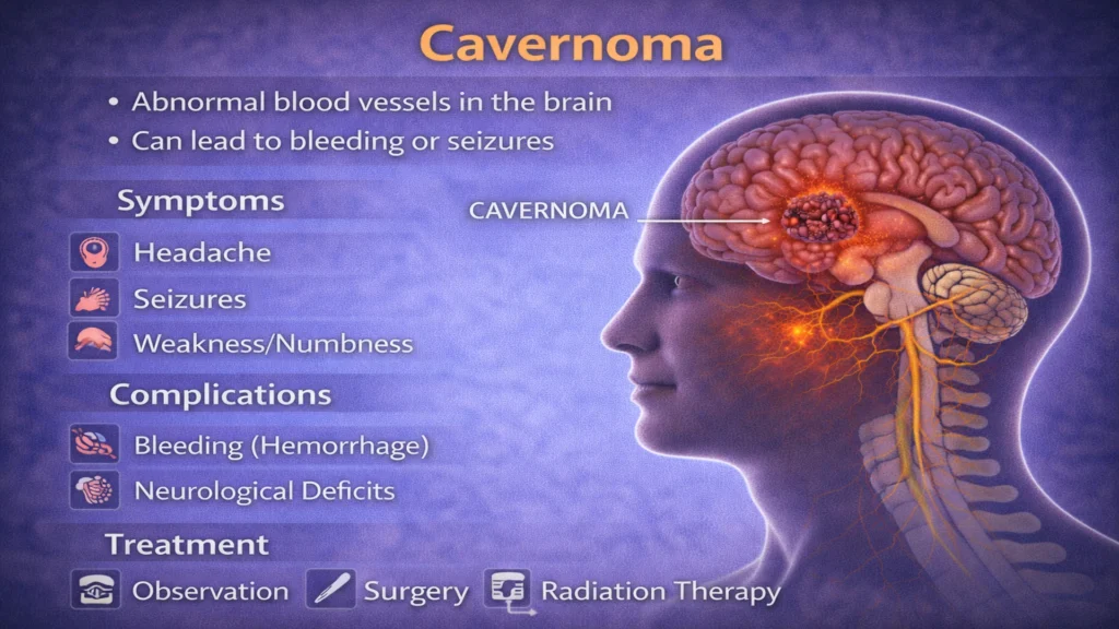
A cavernoma, also known as a cerebral cavernous malformation (CCM), is an abnormal cluster of dilated blood vessels in the brain or spinal cord. These vessels form a lesion with thin walls that are prone to leakage or bleeding.
Cavernomas are not tumors and are usually benign, but their structure makes them potentially dangerous, especially when located in critical areas of the brain.
How a cavernoma forms
Cavernomas may be congenital (present from birth) or develop over time. Some cases are sporadic, while others are associated with inherited genetic conditions.
The malformed vessels inside a cavernoma lack normal structural support, which increases the risk of microbleeds or, in some cases, more significant hemorrhage.
Why cavernomas can be dangerous
The main risk associated with cavernomas is bleeding within the brain tissue. Even small hemorrhages can disrupt normal brain function, depending on the location of the lesion.
Potential dangers include:
- intracerebral hemorrhage
- seizures
- progressive neurological deficits
- recurrent bleeding episodes
Cavernomas located in the brainstem or deep brain structures carry a higher risk due to the sensitivity of these areas.
Common symptoms
Many cavernomas remain asymptomatic for years and are discovered incidentally. When symptoms occur, they may include seizures, headaches, weakness or numbness in the limbs, balance problems, vision changes, or speech difficulties.
Symptoms can appear suddenly if bleeding occurs or develop gradually due to repeated microbleeds.
Diagnosis
Diagnosis is primarily based on MRI of the brain, which can reveal the characteristic appearance of a cavernoma. MRI allows evaluation of lesion size, location, and signs of prior bleeding.
In some cases, additional imaging or neurological testing is used to assess the functional impact of the lesion.
Management and treatment options
Treatment decisions depend on symptoms, lesion location, bleeding history, and overall risk.
Observation
Asymptomatic cavernomas may be monitored with regular imaging and neurological follow-up.
Medical management
Seizures and other symptoms are treated with appropriate medications.
Surgical treatment
Surgical removal may be considered for symptomatic cavernomas, especially after repeated bleeding or when seizures are difficult to control.
Treatment is always individualized, balancing potential benefits and risks.
Prognosis
Many individuals with cavernomas live normal lives, especially when lesions are stable and asymptomatic. Prognosis depends on lesion location, bleeding history, and response to treatment.
With careful monitoring and timely intervention, serious complications can often be prevented or minimized.
When to seek urgent medical attention
Immediate medical evaluation is required if sudden neurological symptoms occur, such as severe headache, weakness, numbness, vision loss, speech difficulties, or seizures. These may indicate bleeding and require prompt assessment.
FAQ — Cavernoma
A cavernoma is an abnormal cluster of fragile blood vessels in the brain or spinal cord.
No. It is a vascular malformation, not a tumor.
Yes. Bleeding is the main risk associated with cavernomas.
No. Many can be safely monitored without surgery.
Yes. Seizures are a common symptom, especially for lesions in the cerebral cortex.
Some forms are inherited, but many cases occur sporadically.


